v.1.09 AH 28.02.2024
Checklist
After your introduction at the Center for Advanced Imaging (CAi), more steps need to be taken before starting to use the microscopes. Do not forget to bring / finish the following documents to the introduction: signed cost & user declaration, the GMO relevant FormblattZ of the GMO used in the introduction and later research and finish the laser safety training and sign it on the declaration.
The checklist is as follows, with instructions below:
- Read the CAi rules
- Complete the laser safety training
- Provide a signed Kostenübernahmeerklärung (cost transfer declaration)
- Provide a project description (template available at CAi website)
- Obtain access to the CAi shared network drive
- Obtain access to the booking system
- Download FIJI / work with microscopy images
CAi rules
Please browse through our short "rules" list, especially to guide you while planning and booking the first microscopy experiments:
http://www.cai.hhu.de/en/service/service/user-rules.html
Laser safety training
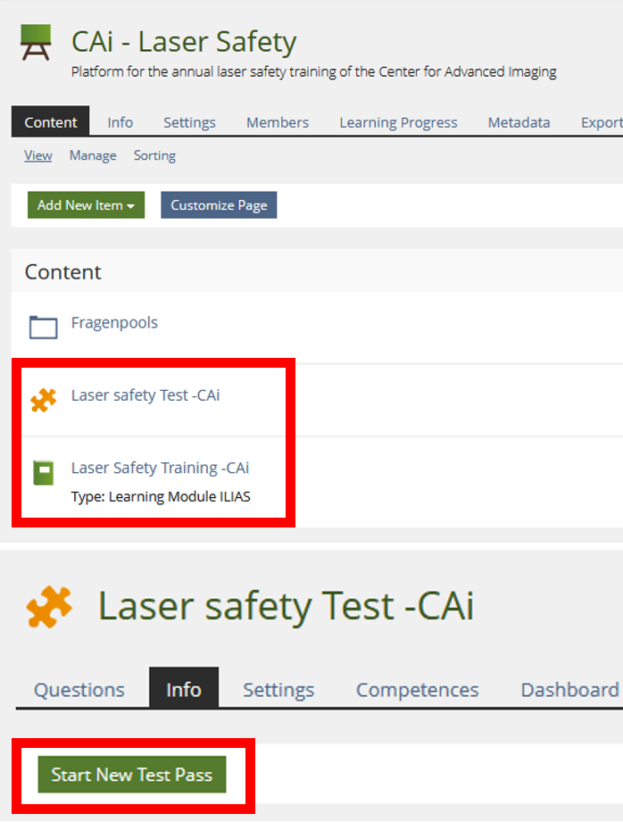
Before your first session at the microscope, you need to complete our short online course on laser safety, and pass the associated quiz. This normally takes about 15-20 minutes.
Location - when opened, please log-in with your university-ID, using “Anmelden” in the top-right.
https://ilias.hhu.de/goto.php?target=crs_489646_rcodeaNYcefCxjk&client_id=UniRZ
Course password: LaserCAi2017

Or use the following links for direct access to the training elements:
Lasersafety training: https://ilias.hhu.de/goto.php?target=lm_1083340&client_id=UniRZ
Pass the Test to complete the training: https://ilias.hhu.de/goto.php?target=tst_1435381&client_id=UniRZ
Kostenübernahmeerklärung
CAi needs a completed cost-transfer declaration ("Kostenübernahmeerklärung”) from you, which has to be signed by your laboratory head, or budget manager.
The form can be downloaded from the bottom of the following page:
http://www.cai.hhu.de/en/service/service/user-costs.html
This confirms the agreement that CAi will charge your microscope costs to the account of your laboratory.
Project description
CAi needs a brief description of your project, specifically describing the use of genetically modified organisms (GMOs) to include: GMO species, strain, DNA constructs/microorganisms used for transformation, antibiotic resistances, tags, promoters, transformation methods and confirmation (PCR, blots etc.), disposal methods, risk group (eg. S1) and the responsible laboratory. Normally, a good existing source for these details is the first pages of the "Formblatt Z" summing up the information about the planned GMO. In addition, there is a template for a project description available at the CAi website (http://www.cai.hhu.de/en/service/service/user-rules.html). Once completed, the project description must be sent to CAi to cover this point. If the type of imaged organism changes over the time of a project (e.g. fixed/ inactivated samples first, live-cell imaging later), this must be reported to the CAi by the user or the PI. The user MUST document in the log-book the name of the GMO, which is matching the name of the detailed information about this GMO of the genetic engineering facility.
Network drive for data storage
After the introduction, CAi will provide your account with access to the central data storage.
For all users, CAi provides a centralized network storage, which is hosted at the ZIM/HHU and is accessible from any computer connected to the HHU network (incl. OpenVPN). This is the main image storage location. Users have access to a private section, that is visible to their laboratory only.
Address: \\cai.ad.hhu.de\daten
Please save your image data on this network drive and not the microscope PC. You may save images locally during your microscopy session, and when done with the session, move all of them to the network drive.
The drive can be opened as follows, also from microscope computers:
- open the file explorer
- right click on "this PC" (or “computer”)
- click on "map network drive" ("Netzlaufwerk verbinden")
- enter the network drive address "\\cai.ad.hhu.de\daten"
- log in using "ad\university-ID" (use your own “Uni-kennung” after ad\)
- move your data (delete from microscope PC)
- disconnect the drive: in “this PC”, right click the network drive and "disconnect"
Mac users, when the above steps are not working, may have to use the following address:
smb://cai.ad.hhu.de/daten
Microscope booking system
After the introduction, you should log onto the CAi booking system for the first time, with your university-ID, and normal password. This automatically creates a new profile for you, which we can give access to the right microscopes. The booking system can be found here:
https://booking-cai.hhu.de/dashboard.php
Working with microscopy images
Commonly, after image acquisition, we use FIJI (ImageJ) to handle the image data. It is recommended to download this program and give it a try: https://fiji.sc/#download. We are happy to help you learn the basic functions in a short session soon after your microscopy introduction. Also, FIJI is open-source, and the internet is full of resources describing FIJI-workflows at any levels of proficiency. CAi furthermore has a broad image analysis tool knowledge and provides general help, advice, and suggestions for image analysis and figure generation.