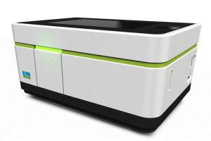
Overview
Service contact (administrators only): "EVO Portal", Perkin Elmer website. |
Incubation: Heating is available (37-42 °C; system temperature of about 25 °C without heating activated). CO2 controller available (1-10 %). No humidity control: Use closed samples in excessive imaging buffer, if necessary.
Objectives: 5x, 20x, 40x (all air).
Light/optics: 4x LED light source (can activate 1 source simultaneously), 5x emission filter (UV / green /yellow / red / far-red), Spinning disc optics. 1x top-down-LED (740 nm, for morphology, using transparent-top samples).
Camera: 4.7 Megapixel sCMOS sensor.
Light path/wavelengths
Excitation windows available:
- 370 nm (355-385 nm)
- 475 nm (460-490 nm)
- 550 nm (530-560 nm)
- 630 nm (615-645 nm)
Detection windows available:
- 430-500 nm (blue)
- 500-550 nm (green)
- 515-580 nm (yellow)
- 570-650 nm (red)
- 665-760 nm (far-red)
What about my specific excitation line?
For fluorophores with an excitation peak at for example 510, use the closest available light source: 475 nm
See https://searchlight.semrock.com/ for excitation/emission spectra of many fluorescent dyes and proteins. When testing a new assay, make sure to include enough controls to verify that no crosstalk appears. |
Plates
Usage
Machine start
- Depress button (green) on the left side of the machine
- Wait a few minutes until the software displays "device is ready".
- The machine is now ready to use
Around the sample area
- If the table is not powered, eject/re-insert he plate holder (software). Do not manually move table.
- Plate/sample holder is mounted on steel tracks: DO NOT CLEAN/TOUCH (if dirty, call service)
- Plate/sample holder contains an internal "penta-pattern" of dots, used for high-accuracy positioning of the sample.
- Red dot on objectives needs to match the red dot on the microscope. Objectives can be manually lifted out of the system.
- With open lid, the machine counts as a class 3B laser. Close the door firmly.
- Emergency release hatch button (lid open) is on the lower right of the machine.
Plate loading
- Start Harmony software
- Log-in with user account: personal account, with personal settings
- User accounts can only delete own data. Administrators can delete all.
- Choose plate. Nomenclature = number of wells, manufacturer, type/name of plate
- Choose objective. Start at eg. 20x / 0.4 NA
- Choose operational mode (opt-mode). Confocal = spinning disc mode, with pinholes, 5% transmission of light, both ways. Epi = non-confocal, 100% transmission of light, both ways.
- Binning: (makes image size <4.7 Megapixel) → signal x4, noise x2, resolution significantly reduced.
- Live preview: eg. Show current image during stack acquisition. off = evaluate manually after setting up acquisition.
- Click "new" → new experiment.
- Set up individual channels for your fluorophores.
Arrowheads
Located under menus always reveal more detailed settings. |
- Plate layout (right-most). Select wells to measure, click "SELECT". Select FOV, number, position.
- Select well-to-test.
- Clean plate-bottom with wipe (KimTech or similar) and 70% Ethanol before use.
- Load plate with or without lid (place within protrusions).
- or: Load slides in holder.
- or: Load slide holder into Operetta.
Setting up for imaging
- Set up the desired channels, select a well, and take a snapshot. Different z-heights may need to be trialled.
- Evaluate brightness; auto-scale and auto-contrast are always applied.
- No range-indicator (over/under exposure) is available, but a histogram for each channel is displayed.
- Histogram: if not visible, can be enabled using the arrow under "Channels" to the right of the image.
- Scaling: Normal mode: Black-to-Colour. Enhanced mode: Black-to-Colour-to-White.
- Right-click on the sample image, "show intensity". Ctrl + Click to add ROIs.
- Find focus height: Z-stack! "test" is used here, and only applies to the well that was highlighted by clicking, on the right.
- Start at negative values. Go up to positive. See in "TEST IMAGES" (right of screen)
- Pick the right focus height for each channel individually.
- Next, to be certain, do focus finding in another cell. Possible plate-tilting may be revealed, and needs inspection.
Z-stacks yes/no?
It is possible to disable "use z-stack in test", to test with only single-plane images
If you want no stack acquisition upon running the actual experiment, simply select "0 planes" "Selecting" wells and highlighting wells
Clicking on wells highlights them, and points at wells to be used in test images
Selecting wells (highlighting, then clicking "select") determines which wells to use in the experiment. |
- Overview picture: in the "well layout" section, select all interesting wells; right click "overview;" preview field: observe the stitched image afterwards.
- The overview picture is automatically downscaled to 200 MB in size; full-resolution stitching is not supported.
- Double-check overlap: Pull up the contrast to an extreme value (visually; right-click in the image)
- Determine whether flat-field correction will be necessary.
- "online-job" = perform image analysis during acquisition
- You can add layouts (labels) to the output data. This is an additional table.
- Re-use a plate layout (saved, from eg. editor or previous experiment); "Assay" in "define layout" (right-most).
Use overview image to pick interesting regions
Using "Test Images", and marking regions, right-click and "use as background for well" for a visual aid for selecting further regions. |
Run the experiment
Rule of thumb for estimated dataset size
The Operetta CLS is only displaying the total size of a dataset if not enough disk space is available. If you want to know the estimated dataset size before you start an experiment, use the following rule of thumb: Number of wells x fields x channels x z-planes x timepoints x 8 MB = approximate dataset size in MB |
Flat-field correction
The software can do flat-field correction based on acquired images, provided that foreground/background and areas without cells are present.
This also works in wells filled with tissue, or more-than-confluent cell cultures. |
- When done: eject the sample (settings - Operetta CLS - eject) and remove plate/slide holder. Afterwards "eject" once more to close the system again.
Exporting data
- In the "settings" menu up top, data management can be used to export data
- Export your data as an "archive" for later re-analysis using Harmony.
- Export your data as "measurement + associated data" to save results as TIFF files, alongside already-performed measurement outcomes, and re-analysis eg. using CellProfiler. Be aware that the exported raw images are not flat-field corrected, so it might be necessary to correct the images before the analysis.
- Connect to the CAi shared network drive to store the data, or bring your own data-carrier (USB).
- After the session, delete your data from the Operetta computer.
Saved parameters
During an experiment, you define and save the following objects: - Experiment (light, stacks, channels, etc)
- Assay layout (cells, treatments, rows/column names)
- Measurement (image data)
- Analysis sequence (processing steps, calculations, values of interest)
- Evaluation (grouping, graphs/charts, heatmaps, well-display, statistics, data points)
|
Image processing and analysis

Loose info pieces:
Z-steps always > 500 nm
Fluorescent staining strategy:
To facilitate auto-detection in subsequent image analysis steps:
a) stain cells with target marker
b) stain cytosol separately (eg. CellMask Blue)
c) stain nuclei separately
Computer attached to the microscope:
DO NOT SWITCH OFF!
DO NOT UPDATE WINDOWS!
The PC is running a live image data and image analysis server.


